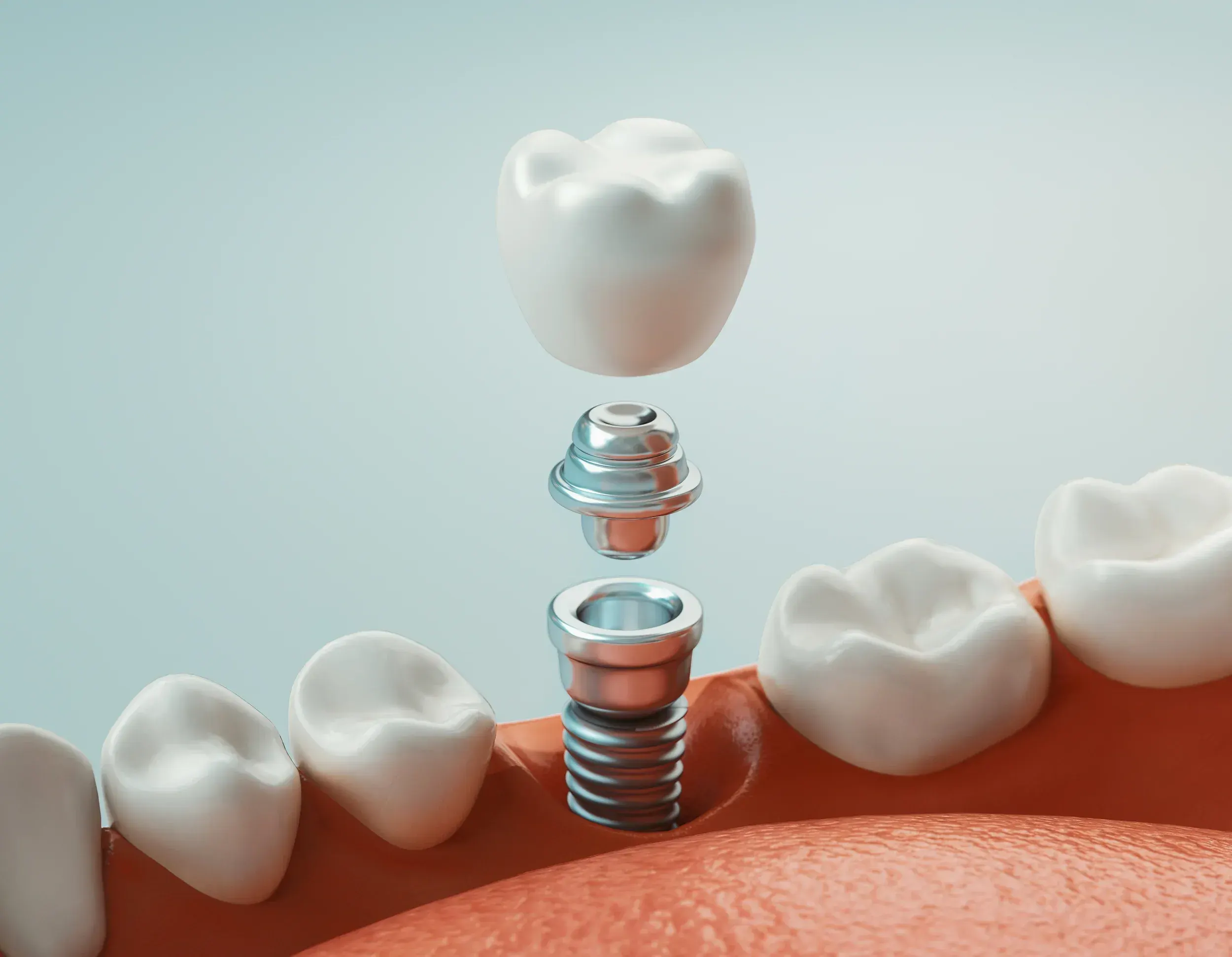
Cavities Between Teeth
Detects interproximal decay that visual exams cannot see


Preventive and General Dentistry
Safe, digital imaging for accurate diagnosis and early detection.
Seeing What the Eye Cannot
Many dental problems start silently. Cavities can hide between teeth, infections can simmer at the root of a tooth, and bone loss from gum disease can progress for years before you notice loose teeth or pain. Wisdom teeth can press against neighbors below the gumline, and small cysts can form without any outward sign. By the time symptoms appear, treatment is often more involved, more expensive, and more uncomfortable than it would have been if the problem were caught early.
Dental X-rays give us a clear view of the structures hidden beneath your gums and inside your teeth. With digital imaging, our team can detect early decay between teeth, evaluate bone levels, monitor existing fillings and crowns, plan implants safely, and track development in children. Because images appear on screen instantly, your dentist can review findings with you in the same visit, explain what we see, and recommend the most conservative treatment available.
The Science of Digital Imaging
Dental X-rays, also called dental radiographs, are diagnostic images that use very small amounts of ionizing radiation to capture pictures of your teeth, gums, jaw, and surrounding structures. According to the American Dental Association, X-rays are an essential diagnostic tool that helps dentists identify problems that cannot be seen during a standard comprehensive dental exam.
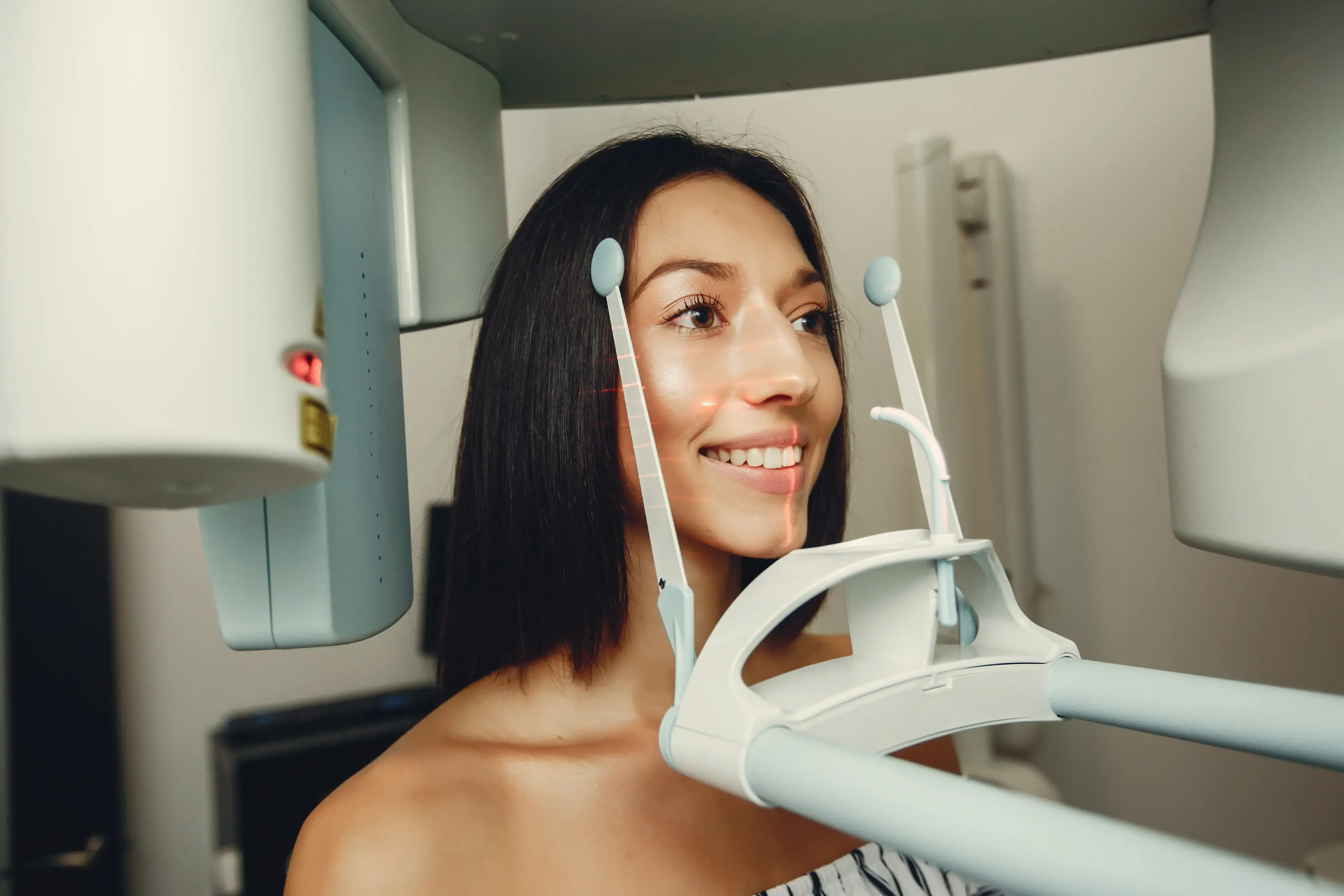
A focused beam of low-dose radiation passes through the mouth and is captured by a digital sensor placed inside the cheek or by a panoramic machine that rotates around the head. Dense tissues like enamel and bone absorb more radiation and appear white, while softer tissues and decay absorb less and appear darker. The FDA notes that modern digital systems can reduce radiation by up to 80 percent compared with traditional film X-rays.
You will be draped with a lead apron and thyroid collar for protection. A small sensor is placed in your mouth or, for a panoramic image, you stand still while the machine circles your head. Each image takes only seconds, and the entire series is typically complete in under 10 minutes.
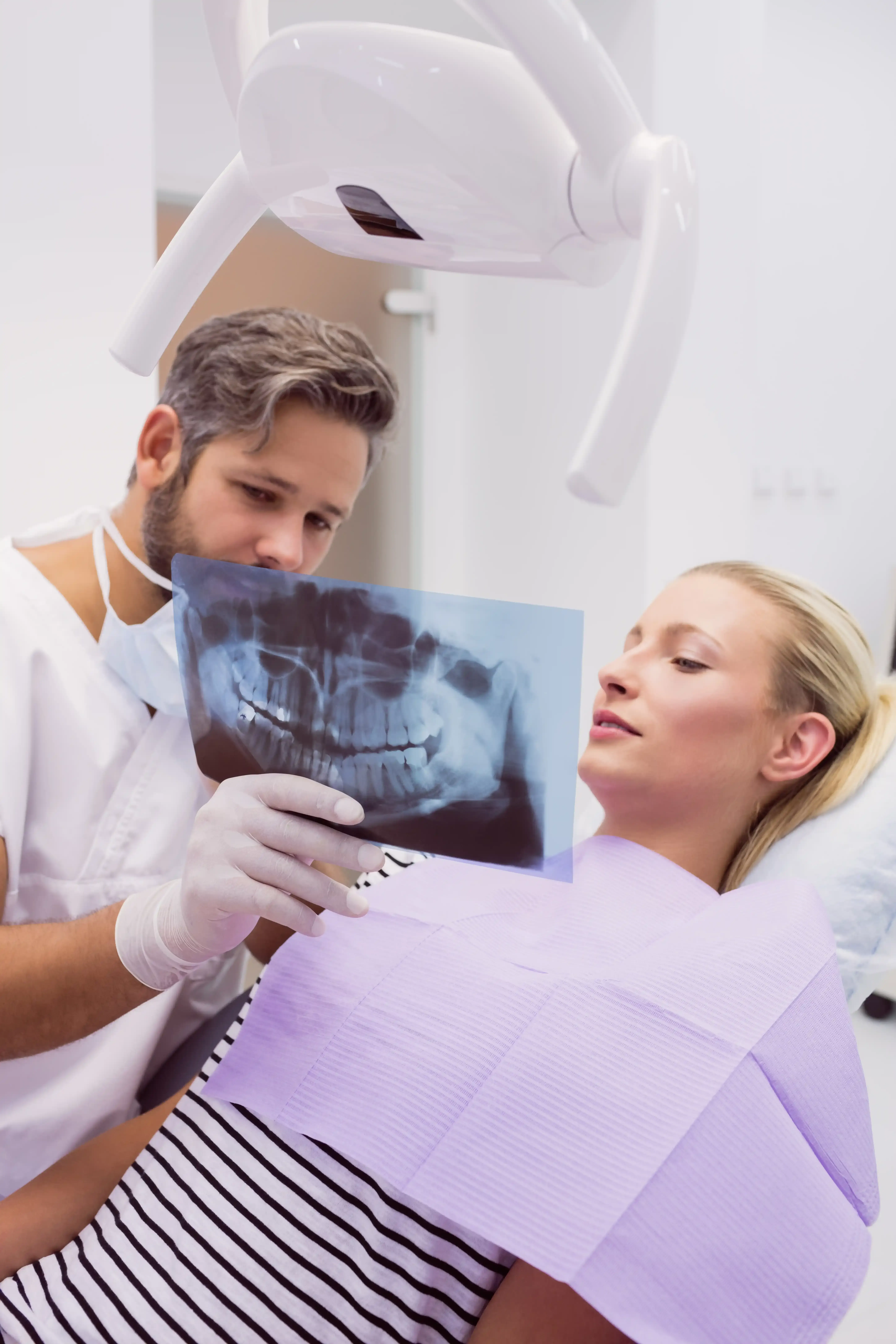
Images appear on the operatory screen within seconds. Your dentist reviews them with you, points out any areas of concern, and discusses next steps in the same appointment, which means no waiting and no return visits to learn results.
Digital sensors use up to 80 percent less radiation than film
Images appear on screen within seconds for same-visit review
Finds decay, infection, and bone loss before pain develops
No chemical processing or film waste, fully digital workflow
Images can be securely sent to specialists or insurance
Typically covered by Delta Dental and most major plans
Compare Your Options
| Treatment | Mechanism | Time | Results | Duration | Downtime | Best For |
|---|---|---|---|---|---|---|
| Bitewing X-rays | Side view of upper and lower teeth | 2-3 minutes | Instant on screen | Used at recall visits | None | Cavities between teeth |
| Periapical X-rays | Full tooth from crown to root tip | 1-2 minutes per image | Instant on screen | Targeted per tooth | None | Root, pulp, abscess evaluation |
| Panoramic X-rays | Single rotating image of entire mouth | Under 20 seconds | Instant on screen | Every 3-5 years typical | None | Wisdom teeth, jaw, full overview |
| 3D Digital Scanning | Cone-beam volumetric imaging | 10-40 seconds | Instant 3D model | Procedure planning | None | Implants, complex anatomy |
Finding Your Best Diagnostic Plan
Most patients benefit from periodic dental X-rays as part of routine preventive care. Frequency depends on your age, oral health history, and current risk for decay or gum disease, following the ADA guidelines for radiographic examinations. Your dentist personalizes your imaging schedule rather than applying a one-size-fits-all approach.
Our team follows the ALARA principle, As Low As Reasonably Achievable, and provides a lead apron and thyroid collar for every X-ray. If you are pregnant or think you may be, tell our team so we can modify or postpone imaging when appropriate.
Your dentist reviews your history, current symptoms, and any prior images you can share.
Our team drapes a lead apron and thyroid collar over you to shield non-target areas.
A small digital sensor is placed in your mouth, or you stand at the panoramic machine.
Each digital image is captured in under a second using a low-dose pulse of radiation.
Images appear instantly on screen, and your dentist walks you through every finding.
What to Know
Most patients experience no side effects at all. Some find the sensor placement briefly uncomfortable, especially if they have a strong gag reflex or a small mouth. Any discomfort resolves the moment the sensor is removed, typically within seconds.
True complications are extremely rare. The amount of radiation from a full set of digital bitewing X-rays is approximately 0.005 millisieverts, which is less than one day of background radiation from the natural environment, according to the FDA radiation safety guidance. Pregnant patients should inform the team so imaging can be postponed or modified when appropriate.
Our practice follows the ALARA principle, As Low As Reasonably Achievable, and uses digital sensors that reduce exposure by up to 80 percent versus traditional film. Lead aprons and thyroid collars are used for every patient. Our doctors and team are trained in current radiation safety protocols and order only the images needed for accurate diagnosis.
Dental X-rays are typically covered by dental insurance as part of routine preventive care, particularly bitewing and periapical images taken during recall visits. Out-of-pocket cost in the East Ellijay and Ellijay area generally ranges from about 25 to 75 dollars for a single image and 90 to 200 dollars for a full mouth or panoramic series, depending on the type and number of images. Your final cost will depend on your insurance plan, the type of X-rays ordered, and any prior images on file. Exact pricing will be discussed during your visit based on your individual treatment plan.
Our office is in-network with Delta Dental, and we work with most major dental insurance plans to maximize your benefits. Many preventive X-rays are covered at 80 to 100 percent depending on your policy.
For any portion not covered by insurance, we offer several financing options to keep care affordable.
If financing helps you move forward with care, our team will explain your options and help you choose the plan that fits your budget.
East Ellijay's Trusted Provider
Three DMD providers including Dr. Settel with 17 years of practice
Up to 80 percent less radiation than traditional film systems
Images reviewed with you in the same appointment, no waiting
Delta Dental in-network, CareCredit and Cherry financing available
Pair your X-rays with these complementary preventive and diagnostic services.
Your Questions Answered
Frequency depends on your age, risk factors, and oral health history. Most healthy adults need bitewing X-rays every 12 to 36 months, while a full series or panoramic image is taken every 3 to 5 years. Your dentist personalizes the schedule to your needs.
Yes. Digital X-rays use very low doses of radiation, up to 80 percent less than traditional film. We follow the ALARA principle and provide a lead apron and thyroid collar for every patient. The exposure from a routine bitewing series is less than one day of natural background radiation.
X-rays show cavities between teeth, bone loss from gum disease, abscesses at tooth roots, impacted wisdom teeth, the condition of fillings and crowns, and developing teeth in children. They reveal problems long before they cause pain or become visible.
Routine X-rays are usually postponed during pregnancy, especially in the first trimester, unless imaging is medically necessary for diagnosis. If you are or might be pregnant, please tell our team before your appointment so we can plan appropriately.
X-rays themselves are painless. Some patients find the sensor placement briefly awkward, especially with a strong gag reflex. Our team uses small sensors and positions them gently. Each image takes only seconds, and the full series is usually complete in under 10 minutes.
Yes, in most cases. If you have recent X-rays from another dentist, share them with us before your visit. Many can be reviewed instead of repeated, which helps reduce your overall exposure and saves time during your appointment.
Most dental insurance plans, including Delta Dental, cover routine bitewing and periapical X-rays as part of preventive care, often at 80 to 100 percent. Panoramic and 3D imaging may have different coverage. Our team will verify your benefits before your visit.